Emergency
Whats app Book an Appointment Second Opinion Second Opinion

Radiology And Imagology

Home Radiology And Imagology

Department of Radiology and Imagology

The Department of Radiology and Medical Imaging at SJAS SUPER SPECIALTY HOSPITAL is a highly specialized, round the clock, full service department that strives to meet all patient and clinician needs in Diagnostic Imaging and Image guided therapies. The department is recognized for providing patients and clinicians, the most advanced radiology and medical imaging services. Our Radiologists strive to provide fellow physicians and surgeons with the tools and vital information they need to achieve diagnostic and therapeutic excellence. In a short span of time since its inception the department of Radiology at SJAS SUPERSPECIALTY HOSPITAL has earned a reputation as being a premier centre in providing imaging and minimally invasive therapeutic services. It caters not only to patients from within the hospital but also to the patients from other medical centres across the state. Services and programs provided at the SJAS SUPER SPECIALTY HOSPITAL Department of Radiology include:

Interventional Radiology

Medical Imaging

Musculoskeletal Imaging

Neuroradiology

Radiology and Imagology

Treatments & Procedures

The department's clinical services are outstanding and SJAS SUPER SPECIALTY HOSPITAL’s Radiology Department's faculty and staff perform thousands of patient examinations, giving patients access to a wide spectrum of radiology services. The department of radiology is manned by an intensively trained team managed by highly qualified and experienced radiologists. It is equipped with technologically advanced equipment and offers a wide range of services, from routine radiography services like X-Ray to specialized Ultrasound, Colour Doppler and CT scan facilities.

Body Imaging

As specialists in the Computed Tomography SJAS SUPER SPECIALTY HOSPITAL’s Department of Radiology conducts CT studies of the chest, abdomen and pelvis, performing CT angiography, CT arterial portography and image-guided biopsy studies. The Body CT Section offers state-of-the-art, multi-detector CT imaging for all clinical indications within the abdomen and pelvis. The service oversees the smooth and safe operation of the modality in the performance of examinations pertaining to neuroradiology, thoracic, cardiovascular, and musculoskeletal imaging. It also coordinates the availability of the scanners in the performance of CT-guided procedures by members of the departmental staff.

Gastrointestinal Imaging

Radiologists in the Gastrointestinal (GI) Radiology Division perform diagnostic imaging studies of the GI tract. The GI Division faculty and staff work closely with gastroenterologists, surgeons, internists and other specialists to evaluate and perform studies on patients with known or suspected GI diseases including:

  • Esophagrams (barium swallows) – for patients with difficulty swallowing or reflux symptoms.
  • Upper GI studies – for patients with dyspepsia, epigastric pain, vomiting or other upper GI conditions.
  • Small bowel follow through studies and small bowel enemas (in which barium is injected through a tube placed in the proximal small bowel) for abdominal pain or diarrhea
  • Barium enemas – for patients with rectal bleeding, colonic symptoms or colon cancer screening.
  •  

Magnetic Resonance imaging (MRI)    ACS 3.0T

The uMR® 780 ushers in a new era of rapid imaging with ACS (AI-assisted Compressed), a deep learning-based MR acceleration technology capable of doing acquisitions in seconds. Featuring exceptional homogeneity and gradient performance, combined with a comprehensive suite of advanced applications, the uMR® 780 enables you to achieve new levels of performance in anatomic and functional imaging.

Mr Spectroscopy

Is a medical imaging technique used in radiology to visualize internal structures of the body in detail. MRI makes use of the property of nuclear magnetic resonance (NMR) to image nuclei of atoms inside the body.

Tractography

A technique called high-definition fiber tractography (HDFT) provides a powerful new tool for tracing the course of nerve fiber connections within the brain -- with the potential to improve the accuracy of neurosurgical planning and to advance scientific understanding of the brain's structural and functional networks.

Functional Imaging

Functional imaging is the study of human brain function based on analysis of data acquired using brain imaging modalities such as functional Magnetic Resonance Imaging (FMRI). The aim is to understand how the brain works, in terms of its physiology, functional architecture and dynamics.

Magnetic resonance imaging (MRI) is a non-invasive test doctors use to diagnose medical conditions.

MRI uses a powerful magnetic field, radiofrequency pulses, and a computer to produce detailed pictures of internal body structures. MRI does not use radiation (x-rays).

Detailed MR images allow doctors to examine the body and detect disease.

Functional magnetic resonance imaging (fMRI) uses MR imaging to measure the tiny changes in blood flow that take place when a certain part of your brain is working.

Cardiology

Comprehensive cardiac package consists of excellent scan acceleration and optimized k-space filling, is provided to address complicated cardiology cases.

Interventional, Neurovascular, Peripheral Vascular And Non Vascular Procedure

Are performed on the advanced Cath Lab with 3D rotational angio facility. All complex neuro vascular procedures are performed. SJAS SUPER SPECIALTY HOSPITAL Radiology Department is one of the very few centres in the country where Varicose Veins of lower limbs are successfully treated with Endovenous laser ablation and Sclerosant foam injection. This is an outpatient procedure done under local anesthesia.

Radiology and Imagology

Technology

The equipment is state of the art at SJAS SUPER SPECIALTY HOSPITAL, in imaging technology, with the latest digital picture archiving and Information Management Systems. The consultants have received extensive training and gained immense experience and expertise in their specific areas of clinical service, which assures consistent, high quality patient care while dealing with rare and complex pathologies.
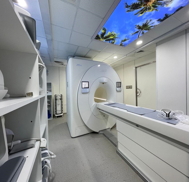
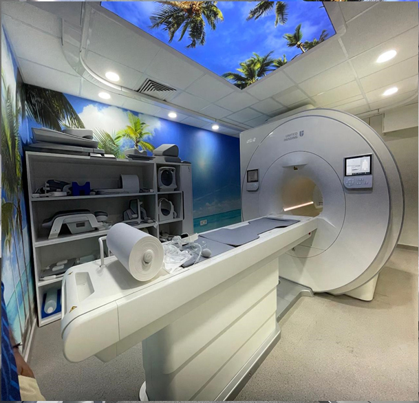
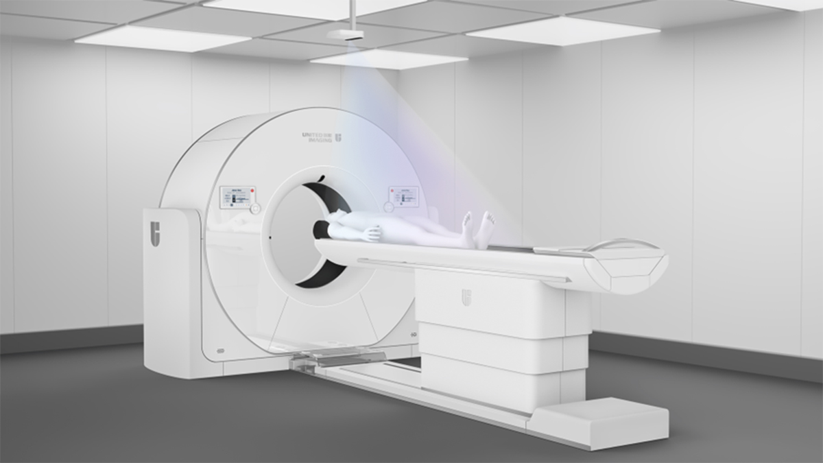
ACS 3.0T MR

The uMR® 780 ushers in a new era of rapid imaging with ACS (AI-assisted Compressed), a deep learning-based MR acceleration technology capable of doing acquisitions in seconds. Featuring exceptional homogeneity and gradient performance, combined with a comprehensive suite of advanced applications, the uMR® 780 enables you to achieve new levels of performance in anatomic and functional imaging..

Break Through the Limits of Speed with ACS (AI-assisted Compressed Sensing)
Intelligent Workflow for Improved Efficiency and Consistency
A Full Range of Advanced Applications for Clinical and Scientific Research
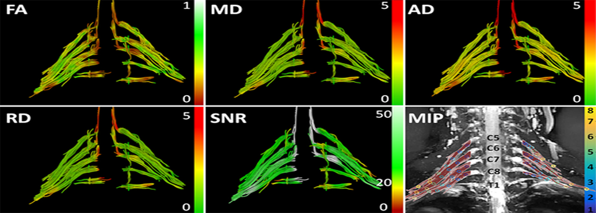
BRACHAIL PLEXUS

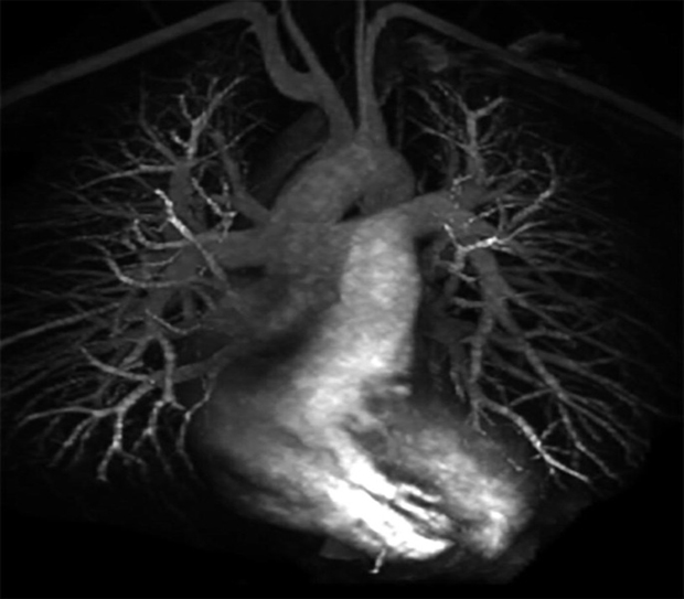
640 DETECTOR ROW MULTI- SLICE CT

The uCT ATLAS is an ultra-premium CT scanner with z-axis coverage of up to 16 cm and up to 640-slices per rotation. The uCT ATLAS features a 0.25s rotation speed, an 82 cm ultra-wide bore and 700 lbs. table weight capacity to accommodate all types of patients, including bariatric. Born with AI, this system features the uAI® Vision 3D camera and utilizes industry-leading AI-empowered technologies throughout the system, offering precise imaging and ease of use for routine to advanced applications, including cardiac, acute care and trauma.

ADVANCED COLOUR DOPPLER AND ULTRASOUND IMAGING

Advanced Colour Doppler and Ultrasound Equipment with Tru Scan Imaging Technology is provided with 3 D & 4 D capabilities and extended field of view. Entire range of Ultra Sonographic examinations like abdomen OB/GYN small parts, vascular, urology, paediatric imaging and various ultrasound guided procedures can be performed with ease.

 DIGITAL RADIOGRAPHY

Digital radiography is a form of radiography that uses x-ray–sensitive plates to directly capture data during the patient examination, immediately transferring it to a computer system without the use of an intermediate cassette.
uDR 780i is a powerful ceiling-mounted x-ray system
Our DR system is equipped with uVision remote technology, which integrates the virtual detector profile, remote collimation and stitching range adjustment functionalities. United Imaging is leading X-ray imaging into a new era by enabling cross-room control, real-time patient video monitoring, and an accelerated exam workflow in an ever-changing clinical environment.